You arrive to a family in winter. Several of them are complaining of headache. A couple feel nauseated. One is more confused than the others. The house smells a little off or maybe it does not smell like anything at all. Everyone’s SpO2 reads 98 to 100 percent.
This is carbon monoxide poisoning until proven otherwise, and your pulse oximeter is the least useful tool you have right now.
Carbon monoxide is called the silent killer for reasons that are directly relevant to prehospital care. It is colorless and odorless. Its clinical presentation mimics dozens of other conditions. And the vital sign monitoring tool that emergency care providers rely on most heavily for respiratory assessment is physiologically incapable of detecting it.
Understanding how and why your monitor fails with CO exposure is the foundation of recognizing this toxidrome before it kills your patient.
Why the SpO2 Reads Normal While the Patient Is Dying
Pulse oximetry works by shining two wavelengths of light through perfused tissue and measuring the ratio of oxyhemoglobin to deoxyhemoglobin based on differential light absorption. It is measuring the saturation of hemoglobin with oxygen.
The problem is that carboxyhemoglobin, hemoglobin bound to carbon monoxide, absorbs light at nearly the same wavelength as oxyhemoglobin. Standard pulse oximeters cannot distinguish between them. When significant amounts of hemoglobin are bound to carbon monoxide instead of oxygen, the oximeter reads those molecules as saturated with oxygen and reports a falsely normal SpO2.
A patient with 40 percent carboxyhemoglobin may display an SpO2 of 99 percent. Their hemoglobin is bound to a molecule that will not offload to tissues. They are functionally hypoxic despite what the monitor says.
This is not a monitoring limitation you can work around by watching the number more closely. The standard pulse oximeter is structurally incapable of detecting CO poisoning. Recognizing the syndrome requires clinical assessment and, where available, specialized monitoring.
What Carbon Monoxide Actually Does
Carbon monoxide binds to hemoglobin with approximately 250 times the affinity of oxygen. Once bound, it holds tightly and does not release to tissues. The hemoglobin carrying CO is functionally useless for oxygen transport, and the body’s tissues are progressively starved of oxygen despite what the pulse oximeter reports.
The additional mechanism that makes CO particularly dangerous is its effect on the oxygen dissociation curve. Even the hemoglobin that is not bound to CO offloads oxygen less effectively in the presence of carboxyhemoglobin. The shift in the dissociation curve means that even residual oxygen-carrying hemoglobin delivers less oxygen to tissues than it normally would.
At the cellular level, CO also binds to cytochrome c oxidase in the mitochondria, directly disrupting cellular respiration. Even if you could get oxygen to the tissues, the cells would have difficulty using it.
The result is a form of cellular hypoxia that does not announce itself on the monitors most providers rely on and that affects every organ system simultaneously.
Who Gets It and When
Carbon monoxide poisoning has a distinct seasonal and situational pattern that prehospital providers can use as a pre-arrival filter.
Winter presentations: The beginning of heating season is peak CO poisoning season. Furnaces that have been dormant through warm months may have cracked heat exchangers, blocked flue pipes, or other defects that produce CO and vent it into living spaces. The first cold day of the year, when furnaces are turned on for the first time, is the highest-risk day for CO exposure from heating equipment.
Multiple patients in the same space: CO poisoning is one of the few toxidromes that predictably affects multiple people in the same environment simultaneously. A call for a family or group with similar vague complaints including headache, dizziness, nausea, fatigue, or altered mental status in a shared indoor space should generate CO as a top differential regardless of season.
Enclosed combustion sources: Generators run indoors during power outages are a leading cause of CO poisoning deaths. Charcoal grills used inside during winter, vehicles left running in attached garages, and gas-powered equipment operated in enclosed spaces all produce lethal CO concentrations rapidly.
Intentional exposure: Carbon monoxide is also used in deliberate self-harm, most commonly by running a vehicle in an enclosed space. Any patient found unresponsive in or near a vehicle in a closed garage should be assumed CO-exposed until proven otherwise.
In the absence of specialized monitoring, CO poisoning is a clinical and contextual diagnosis.
Symptoms range by exposure level: At lower levels, headache, dizziness, nausea, and fatigue predominate. These are symptoms that patients and providers alike attribute to influenza, food poisoning, or stress, which is why CO poisoning is frequently misdiagnosed. As levels rise, confusion, visual disturbances, chest pain, and syncope appear. At high levels, seizures, coma, and cardiac arrest occur.
The classic cherry-red skin: Severely elevated carboxyhemoglobin produces bright cherry-red discoloration of the skin and mucous membranes due to the color of carboxyhemoglobin. This finding is reliable but late, typically appearing at carboxyhemoglobin levels associated with severe toxicity or death. Waiting for cherry-red skin before considering CO poisoning is waiting too long.
The SpO2 discrepancy: A patient who appears clinically unwell, is tachycardic or dyspneic, or is altered, with a normal or near-normal SpO2, in a contextually appropriate setting is suspicious for CO poisoning. The discrepancy between clinical appearance and pulse oximetry reading is itself a diagnostic clue.
Specialized monitoring: CO-oximeters, which measure carboxyhemoglobin directly, can be incorporated into some prehospital monitoring systems. The RAD-57 pulse CO-oximeter is one device used in EMS settings to measure SpCO non-invasively. If your agency carries this technology, use it on any patient with a presentation suspicious for CO. If you do not have it, the absence of a reading does not rule out CO poisoning.
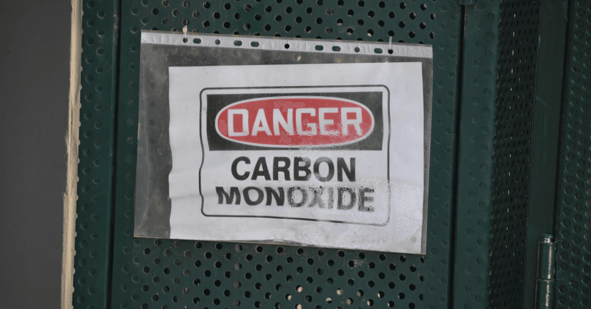
Carbon monoxide is immediately dangerous to life and health at concentrations above 1,200 parts per million. At those concentrations, incapacitation can occur within minutes and death within an hour. You can become the second patient before you recognize what is happening.
Before entering any scene with suspected CO exposure, assess the environment. Personal CO monitors provide a rapid alarm and should be part of the equipment carried by prehospital providers responding to potential CO scenes. If you do not have a personal monitor, your department’s atmospheric monitoring equipment should be used before entry.
If the scene reads positive for CO or cannot be assessed, do not enter without appropriate respiratory protection. A self-contained breathing apparatus provides adequate protection. A surgical mask does not.
Get patients out of the environment before beginning your clinical assessment. Fresh air is the first therapeutic intervention, and it costs nothing if you are already moving the patient to the ambulance.
Remove from the source: Get the patient out of the CO environment and into fresh air as rapidly as possible. Every additional minute of exposure increases carboxyhemoglobin levels.
High-flow oxygen: This is the primary prehospital treatment for CO poisoning. Breathing room air, the half-life of carboxyhemoglobin is approximately four to five hours. Breathing 100 percent oxygen reduces that half-life to approximately 60 to 90 minutes. High-flow oxygen via a tight-fitting non-rebreather mask accelerates the dissociation of CO from hemoglobin.
Airway management for unresponsive patients: A patient who is unresponsive or seizing from CO poisoning needs a definitive airway to ensure reliable oxygen delivery at high concentration.
Cardiac monitoring: CO poisoning can cause myocardial damage and dysrhythmias. Any patient with significant CO exposure should have cardiac monitoring established. A 12-lead is appropriate if time and clinical status allow.
IV access: Establish standard IV access to support medication administration, fluid resuscitation if the patient is hypotensive, and the ability to treat dysrhythmias or other complications that may develop during transport.
When to Think Hyperbaric
Hyperbaric oxygen therapy, which delivers 100 percent oxygen at above-atmospheric pressure, accelerates CO elimination more rapidly than normobaric high-flow oxygen and may reduce the neurological complications of CO poisoning in selected patients.
The indications for hyperbaric referral include loss of consciousness at any point during the exposure, neurological symptoms other than mild headache, carboxyhemoglobin levels above approximately 25 percent, cardiac involvement, and pregnancy due to fetal sensitivity to CO.
Know which facilities in your region have hyperbaric capabilities before the call. Not all hospitals have hyperbaric chambers, and the transport decision may need to be made early in the call when the patient’s condition is still evolving.
References
- Weaver, L.K. (2009). Carbon monoxide poisoning. New England Journal of Medicine, 360(12), 1217-1225. https://doi.org/10.1056/NEJMcp0808891
- Hampson, N.B., et al. (2008). Practice recommendations in the diagnosis, management, and prevention of carbon monoxide poisoning. American Journal of Respiratory and Critical Care Medicine, 177(11), 1179-1184. https://doi.org/10.1164/rccm.200710-1615ST
- Centers for Disease Control and Prevention. (2022). Carbon monoxide poisoning: Fast facts. https://www.cdc.gov/niosh/topics/co/default.html
- Kao, L.W., & Nañagas, K.A. (2004). Carbon monoxide poisoning. Emergency Medicine Clinics of North America, 22(4), 985-1018. https://doi.org/10.1016/j.emc.2004.05.003