First Pass Success Starts Before You Touch the Laryngoscope: A Prehospital Airway Setup Guide
Every failed intubation attempt increases risk. The cords get obscured by blood or secretions from the previous pass. Oxygen saturation drops. The patient's physiological reserve narrows. The provider's stress level rises, and stress degrades fine motor technique. What looked like a manageable airway on the first attempt can become a genuine emergency by the third.
The evidence on first pass success in emergency intubation is consistent: setup matters more than skill, and most preventable failures happen before the laryngoscope ever goes in the mouth. Here is what the research and clinical experience say about building an airway approach that gives you the best possible chance of getting it right the first time.
Why Most Failed Airways Are Not a Procedure Problem
The instinct when an intubation fails is to attribute it to technique. The blade angle was off. The tube was not shaped right. Visualization was inadequate. These are real factors, but they are downstream of a more fundamental set of decisions made before the attempt began.
Positioning was not optimized. The patient was not adequately preoxygenated. The team did not have a clear role structure and something critical was missed. The medication was pushed before the patient was ready. The provider chose to attempt direct laryngoscopy on a patient who needed video. These are setup failures, and they are largely preventable.
No matter how good you are with a laryngoscope, you cannot fully compensate for a patient who is in the wrong position, desaturating before the attempt, or about to go into peri-intubation cardiac arrest because the blood pressure was not addressed first. The procedure is the easy part. The setup is where airway management is actually won or lost.
Know Your Indication Before You Open the Airway Bag
The three primary indications for intubation in the prehospital setting are airway patency, oxygenation and ventilation support, and preemptive airway control for a condition expected to deteriorate. Each of these drives a different clinical context and a different degree of urgency.
Airway patency issues, the patient who cannot protect their airway due to altered mental status, intoxication, head injury, or loss of protective reflexes, represent the classic RSI indication. These patients need a definitive airway, and delay increases aspiration risk and neurological injury.
Oxygenation and ventilation support, the exhausted DKA patient or the late-stage CHF patient working too hard to sustain adequate breathing, requires careful consideration of what happens to the patient's physiology after intubation. A DKA patient is compensating for profound metabolic acidosis by blowing off CO2. Intubating that patient without a plan to match their pre-intubation minute ventilation can cause rapid acid accumulation and cardiovascular collapse. The decision to intubate is correct; the execution requires metabolic awareness.
Preemptive intubation for anticipated deterioration, the burn patient with signs of inhalation injury, the anaphylaxis patient with progressive angioedema, the septic patient heading toward respiratory failure, requires acting before the window closes. These airways become dramatically harder as the underlying pathology progresses. Acting early on a still-manageable airway is consistently better than acting late on an airway that has become a surgical problem.
And the important corollary: not every altered patient needs to be intubated. The postictal patient, the hypoglycemic patient, the overdose patient who may respond to reversal, all of these deserve a brief reassessment before committing to an invasive airway. GCS of eight does not equal intubate. Clinical context does.
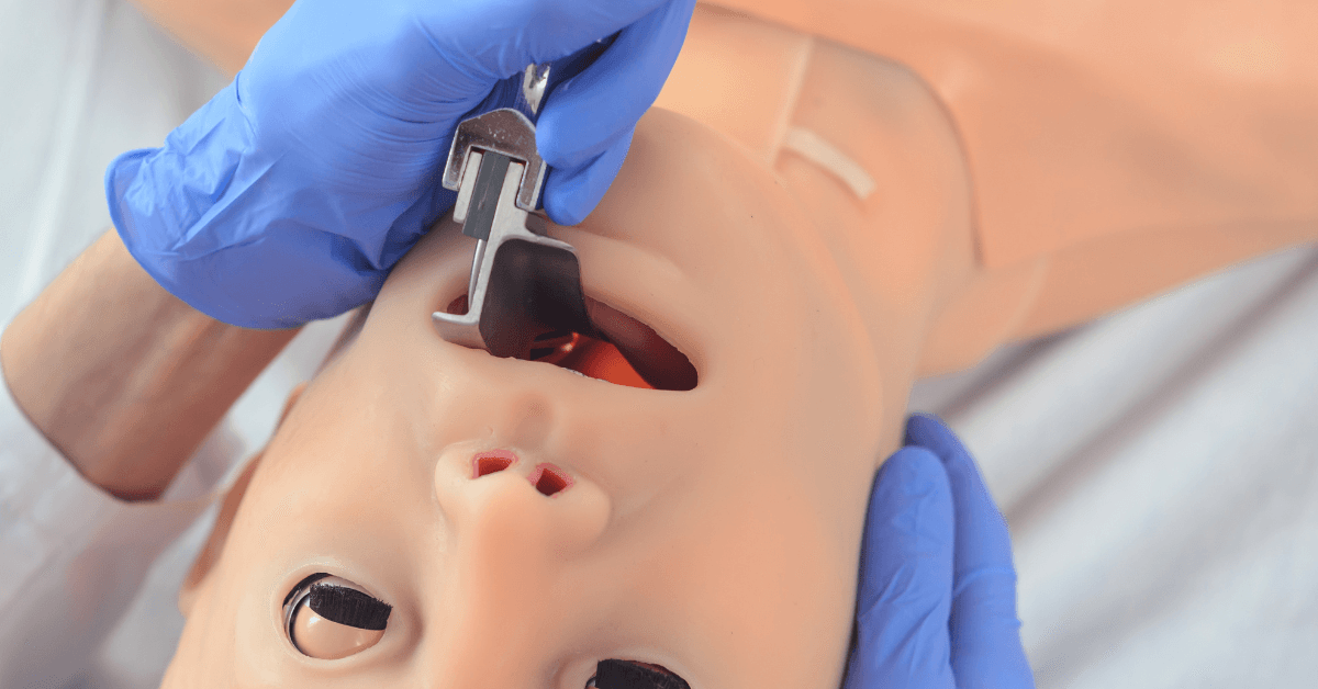
Airway Assessment: The 3-3-2 Rule and What You Are Actually Looking For
Formal airway assessment tools like the Mallampati score exist and have value in the elective surgical setting. In the prehospital environment, you rarely have the luxury of a cooperative seated patient opening their mouth on command. What you do have is a visual and tactile assessment that takes less than 60 seconds and gives you critical information about what you are about to attempt.
The 3-3-2 rule evaluates three anatomical dimensions. Three fingers of mouth opening: less than three fingers suggests limited visualization and reduced instrument maneuverability. Three fingers from chin to hyoid: a short thyromental distance indicates an anterior larynx and a harder corner to make with your tube. Two fingers from hyoid to thyroid notch: a short hyolaryngeal distance suggests limited laryngeal mobility. Any one of these findings alone should raise your index for a difficult airway. Two or more together should change your approach.
Beyond the formal rule, look at the whole patient. Large neck circumference, prominent teeth, limited neck extension, facial hair that will compromise your BVM seal, a beard that will ruin your mask seal before you even get to the tube. These are all factors that compound the difficulty of the attempt and every one of them is visible before you commit.
Mark the cricothyroid membrane with a surgical marker before you begin. It takes 10 seconds and removes one cognitive task from a moment when cognitive load will be at its highest. If you need a surgical airway, you do not want to be palpating anatomy under stress with hands that are not steady.
Positioning Is the Most Underrated Airway Intervention
If you are intubating a patient on the floor, you are making the procedure harder than it needs to be. If you are intubating a patient whose head is perfectly flat without any sniffing position, you are fighting anatomy that you created. Positioning is the single highest-yield intervention in airway management and it costs nothing except the 60 to 90 seconds it takes to do it right.
The goal of positioning is to align three anatomical axes: the oral axis, the pharyngeal axis, and the tracheal axis. When a patient is lying perfectly flat, the oral axis is roughly perpendicular to the tracheal axis, creating approximately a 90-degree angle that makes visualization of the vocal cords and passage of the tube genuinely difficult. The sniffing position, achieved by elevating the head approximately 8 to 10 centimeters with padding while extending the atlanto-occipital joint, brings these axes into closer alignment and dramatically improves your view.
A practical landmark: position the patient so that the tragus of the ear is at the same horizontal level as the sternal notch. With the chin and glabella on the same horizontal plane, you have achieved the sniffing position. This works for most adults. For pediatric patients with proportionally larger heads, padding under the shoulders rather than the head is required to achieve the same alignment. For obese patients, ramping with enough padding to elevate the head and upper thorax 25 to 30 degrees is essential because the weight of thoracic tissue on a flat patient makes both ventilation and visualization significantly harder.
Get the patient off the floor and onto the cot. Adjust the height so you are not bent over at an awkward angle. These seem like obvious steps and they get skipped constantly, especially in cardiac arrest situations where the urgency creates a pressure to act immediately in whatever position the patient happens to be in.
Preoxygenation and Nitrogen Washout: Buying Time Before the Attempt
The goal of preoxygenation before RSI is to replace the nitrogen that makes up approximately 78 percent of room air in the functional residual capacity of the lungs with oxygen. This nitrogen washout creates an oxygen reservoir in the lungs that extends the safe apnea time, the window between paralysis and desaturation, from roughly 1 to 2 minutes on room air to 8 minutes or more with adequate preoxygenation in a healthy adult.
High-flow nasal cannula at 15 liters per minute, combined with a non-rebreather mask, achieves both preoxygenation and passive oxygenation during the apneic period. Leaving the nasal cannula in place during the intubation attempt, even after the patient is paralyzed and apneic, allows continued passive diffusion of oxygen into the alveoli along the concentration gradient. This technique, apneic oxygenation, has been shown in multiple studies to extend safe apnea time and reduce the rate of desaturation during laryngoscopy.
Two to three minutes of high-flow preoxygenation before induction is the standard. If you are pushed for time because the patient is critically ill, even 60 seconds of preoxygenation on a non-rebreather is significantly better than none. Dentures stay in during this phase. They provide facial structure that improves mask seal. Remove them immediately before the intubation attempt.
Team Roles, Checklists, and the Crowd Bias Problem
In the emergency department, a well-run RSI involves a physician managing the airway, a respiratory therapist managing equipment and the ventilator, a pharmacist or nurse drawing and calling out medications, and a charge nurse coordinating the team. Every person has a defined role. The procedure runs smoothly not because everyone is talented but because the system removes ambiguity about who is responsible for what.
In the prehospital environment, you may have two or three people and no checklist. This creates a specific cognitive hazard called crowd bias, the assumption in a group setting that someone else has handled a task. In a team of three people surrounding a patient being intubated, it is entirely possible for the SpO2 probe to not be on the patient when induction meds are pushed, because everyone assumed someone else attached it.
A laminated airway checklist, read aloud by a designated team member who is not performing the intubation, eliminates this failure mode. It should include confirmation that monitoring is attached, IV access is established, the patient's weight has been estimated, drug doses have been calculated, suction is turned on and in hand, backup airway devices are open and accessible, and a contingency plan has been stated. A checklist is not a sign of inexperience. It is what experienced teams use because experienced teams have seen what happens without one.
RSI Sequencing: Sedate First, Always
Rapid sequence intubation involves administering an induction agent followed by a neuromuscular blocking agent in rapid succession to achieve unconsciousness and paralysis for intubation. The critical word in that sequence is followed. The induction agent goes first, every time, without exception.
Administering a paralytic without sedation leaves a fully conscious patient unable to move, breathe, or communicate their distress. This is not a theoretical risk. It has happened in prehospital and emergency department settings when the sequence was rushed or when providers switched the order under pressure. It is one of the most significant harms a provider can cause during airway management and it is entirely preventable.
Push your induction agent, then wait. Give the medication time to take effect. You will see the patient relax. Their jaw will loosen. The reflexes that were protecting the airway will diminish. Then push the paralytic and wait again before attempting laryngoscopy. Giving the medications and immediately reaching for the laryngoscope before either drug has taken full effect means you are attempting intubation in a patient who is partially sedated and partially paralyzed, which is harder than either fully awake or fully down.
Ketamine is an excellent induction agent for most prehospital RSI scenarios because it supports blood pressure, provides analgesia, and can be used for post-intubation sedation from the same vial. Rocuronium has largely replaced succinylcholine as the preferred paralytic in systems where sugammadex is available as a reversal agent. If you cannot get the tube and the patient is rocuronium-paralyzed, sugammadex can reverse the paralysis within minutes and restore spontaneous ventilation.
Resuscitate Before You Intubate
Peri-intubation cardiac arrest is a well-documented complication of RSI in critically ill patients. The mechanism is multifactorial: induction agents cause vasodilation and myocardial depression in a patient who is already hemodynamically compromised, positive pressure ventilation reduces venous return, and the elimination of the patient's endogenous catecholamine-driven compensation creates a sudden hemodynamic cliff.
In a patient with a shock index above 1.0, tachycardia relative to blood pressure indicating significant hemodynamic compromise, the risk of peri-intubation arrest is substantially elevated. The appropriate response is not to rush the intubation but to resuscitate before attempting it. Establish IV access, run fluids, have your vasopressor drawn and ready at the bedside. If the patient's blood pressure is below 90 systolic, get it up before you push induction meds, not after.
Delayed sequence intubation, in which the patient is given a dissociative dose of ketamine to achieve calm and cooperation without full sedation, then preoxygenated for several minutes before paralysis and intubation, is a valuable technique for the agitated hypoxic patient who cannot tolerate a mask and is not cooperating with preoxygenation. It converts a chaotic situation into a controlled one before committing to paralysis.
After the Tube: Confirmation and Post-Intubation Priorities
Tube placement confirmation is not optional and visual confirmation alone is not sufficient. Watching the tube pass the vocal cords is reassuring but does not rule out esophageal intubation, which can occur even under direct visualization in difficult circumstances. The standard of confirmation is waveform capnography showing a consistent, appropriately shaped CO2 waveform over multiple breaths. Bilateral breath sounds and chest rise add supporting evidence but are not substitutes for end-tidal CO2 confirmation.
After confirmation, secure the tube, document the depth at the teeth, and reassess the patient's hemodynamics. Post-intubation hypotension is common and requires immediate attention. Start post-intubation sedation to prevent awareness and manage the ventilated patient's work of breathing. Communicate tube depth, confirmation method, medication doses, and hemodynamic status clearly at handoff.
The Bottom Line
The providers who consistently achieve first pass success in difficult prehospital conditions are not the ones with the most natural talent for laryngoscopy. They are the ones who treat setup as seriously as procedure, who position every patient correctly, who preoxygenate before every attempt, who use checklists to catch what stress makes easy to miss, and who resuscitate before they intubate.
The airway itself is usually three seconds of work. Everything that determines whether those three seconds go well happens in the minutes before.